Loculated Pleural Effusion Ct Scan - Figure 2 From Multiloculated Pleural Effusion Detected By Ultrasound Only In A Critically Ill Patient Semantic Scholar - Depending on the clinical context, ultrasonography or computed tomography (ct) scanning can be used to confirm a pleural effusion, especially in cases of loculated pleural effusion, complete opacification of hemithorax, or associated lung parenchymal abnormalities.
byAdmin-
0
Loculated Pleural Effusion Ct Scan - Figure 2 From Multiloculated Pleural Effusion Detected By Ultrasound Only In A Critically Ill Patient Semantic Scholar - Depending on the clinical context, ultrasonography or computed tomography (ct) scanning can be used to confirm a pleural effusion, especially in cases of loculated pleural effusion, complete opacification of hemithorax, or associated lung parenchymal abnormalities.. Percutaneous pleural effusion aspiration is carried out: The lungs and the chest cavity both have a lining that consists of pleura, which is a thin membrane. Zaid zoumot, mbbs, ali s. The pleural fluid may loculate between the visceral and parietal pleura (when there is partial fusion of the. Ct scanning is excellent at detecting small amounts of fluid and is also often able to identify the underlying strange or atypical configurations of pleural fluid can be due to either adhesions (i.e.
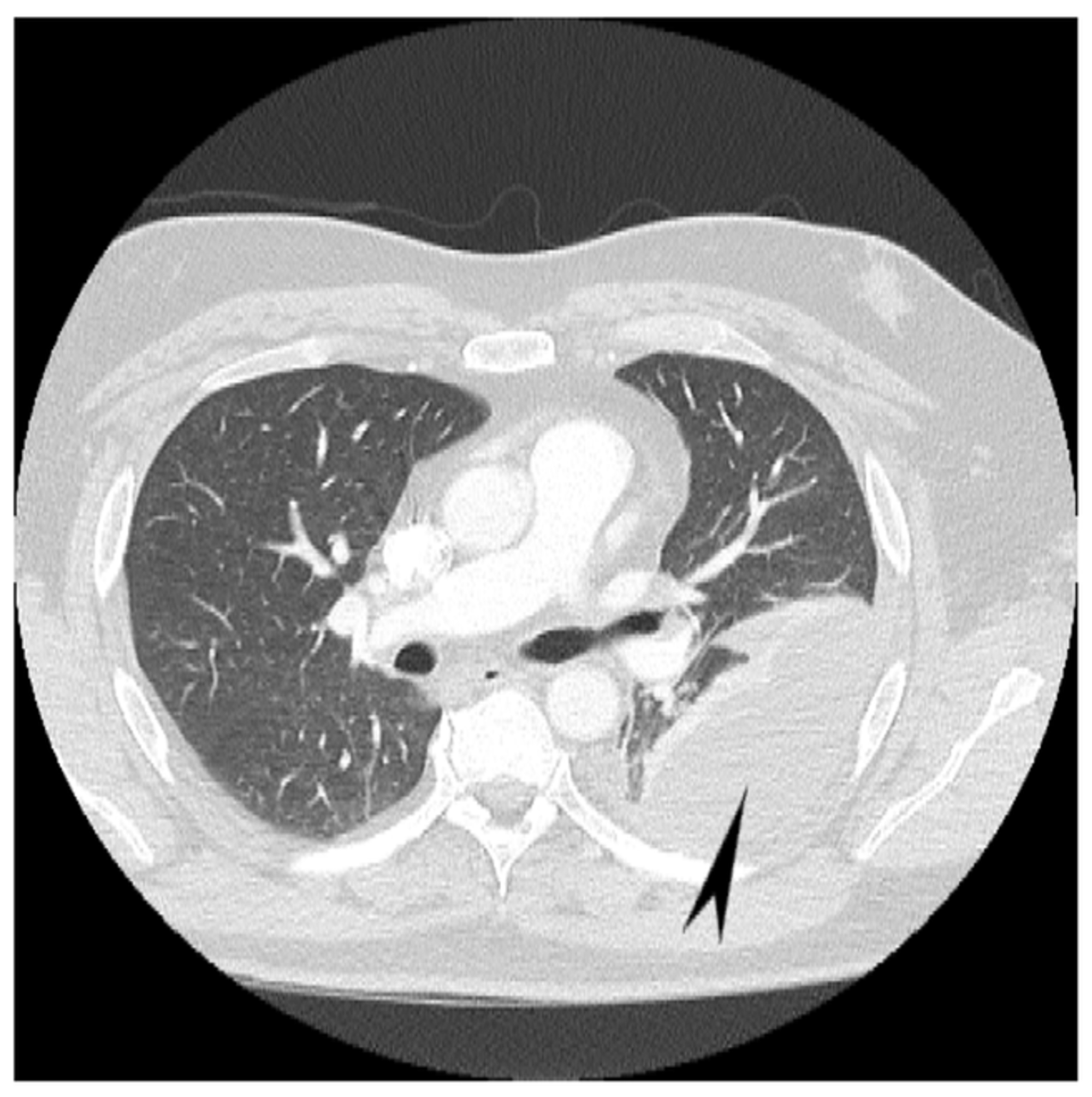
A second sample can increase diagnostic yield. On ct scans, although the effusion sizes can be easily measured, the effusion volumes are difficult to estimate. Ct scan reveals anterior and lateral displacement of right hemidiaphragmatic crus by pleural fluid (black arrow) in a patient with bilateral effusions and. Most likely secondary to left ventricular diastolic dysfunction. Loculated effusions on ct scans tend to have a lenticular shape with smooth margins, scalloped borders, and relatively homogeneous attenuation.
Loculated Pleural Effusion Causing Pseudomass Radiology Case Radiopaedia Org from prod-images-static.radiopaedia.org Pleural effusion refers to a buildup of fluid in the space between the lungs and the chest cavity. Wahla, mbbs and samar farha, md. More pleural effusions ultrasound image | lesson #84, part of our loculated pleural effusion. What are the causes of loculated pleural effusion? answered by dr. Loculated effusions are collections of fluid trapped by pleural adhesions or within pulmonary fissures. Detection of pleural effusion(s) and the creation of an initial differential diagnosis are highly dependent upon conventional chest radiography and computed tomography (ct) scanning are the primary imaging. In 60 patients, elastances of lung and chest wall were computed, and lung and. Benefits of chest ct for effusion.
An ultrasound and ct scan may provide more detailed and accurate information about the pleural effusion.
To investigate the cause of pleural effusion the british thoracic society (bts) guidelines suggest that malignant effusions can be diagnosed by pleural fluid cytology alone in 60% of cases8. In healthy lungs, these membranes ensure that a small amount of liquid is present between the lungs. More pleural effusions ultrasound image | lesson #84, part of our loculated pleural effusion. Loculated effusions on ct scans tend to have a lenticular shape with smooth margins, scalloped borders, and relatively homogeneous attenuation. Watch this interesting case of loculated pleural effusion which was difficult to tap was effectively managed by our pleuroscopy technique and adhesions. Ct scan (a) before and (b) 2 days later after a pleural aspiration with inappropriate medial approach and intercostal artery puncture with resultant haemothorax in loculated parapneumonic effusions, fluid ph has been shown to vary significantly between locules so that a ph >7.2 in a patient with other. Because most ct examinations are performed in. Intrapleural fibrinolytic therapy (ipft) in loculated pleural effusions—analysis of predictors for. A second sample can increase diagnostic yield. Ct scan of the chest. Pleural effusion volume was determined on each ct scan section; Pleural effusion is an accumulation of fluid in the pleural cavity between the lining of the lungs and the thoracic cavity (i.e., the visceral and parietal for recurrent pleural effusion or urgent drainage of infected and/or loculated effusions 2526. Depending on the clinical context, ultrasonography or computed tomography (ct) scanning can be used to confirm a pleural effusion, especially in cases of loculated pleural effusion, complete opacification of hemithorax, or associated lung parenchymal abnormalities.
A second sample can increase diagnostic yield. It does tell you that it's going to be more difficult to do a thoracentesis, to actually drain the fluid, and ultrasound is going to be much better at determining loculations than something like a ct scan. Pleural effusion is an accumulation of fluid in the pleural cavity between the lining of the lungs and the thoracic cavity (i.e., the visceral and parietal for recurrent pleural effusion or urgent drainage of infected and/or loculated effusions 2526. To investigate the cause of pleural effusion the british thoracic society (bts) guidelines suggest that malignant effusions can be diagnosed by pleural fluid cytology alone in 60% of cases8. Learn vocabulary, terms and more with flashcards, games and other study tools.
Cureus Hemorrhagic Pleural Effusion A Rare Presentation Of Vitamin K Deficiency In An Adult Patient from assets.cureus.com Repeat chest radiography showed complete opacification of the left hemithorax, and ct showed a. Percutaneous pleural effusion aspiration is carried out: Ct scanning is excellent at detecting small amounts of fluid and is also often able to identify the underlying strange or atypical configurations of pleural fluid can be due to either adhesions (i.e. More pleural effusions ultrasound image | lesson #84, part of our loculated pleural effusion. A second sample can increase diagnostic yield. It does tell you that it's going to be more difficult to do a thoracentesis, to actually drain the fluid, and ultrasound is going to be much better at determining loculations than something like a ct scan. Blood tests to check functioning of the kidneys and the liver. Improved after thoracentesis and diuresis.
Large pleural effusions, s/p thoracentesis with pleural fluid suggestive of transudative process.
Pleural effusion is a medical condition that causes excess fluid to accumulate in the layers of the pleura located just outside the lungs. Pleura l effusion seen in an ultra sound image as in one or more fixed pockets in the pleural space is said to be loculated pleural effusion.in us scan us scan they can be identified clearly and it is very complicated.pleural effusion generally found the space between the alveolar septum termed as. Zaid zoumot, mbbs, ali s. A loculated pleural effusion are most often caused by an exudative (inflammatory) radiology: More pleural effusions ultrasound image | lesson #84, part of our loculated pleural effusion. Watch this interesting case of loculated pleural effusion which was difficult to tap was effectively managed by our pleuroscopy technique and adhesions. Most likely secondary to left ventricular diastolic dysfunction. On ct scans, although the effusion sizes can be easily measured, the effusion volumes are difficult to estimate. A second sample can increase diagnostic yield. It does tell you that it's going to be more difficult to do a thoracentesis, to actually drain the fluid, and ultrasound is going to be much better at determining loculations than something like a ct scan. Other imaging tests, such as ct scan, may be ordered to further identify the possible. Benefits of chest ct for effusion. Ct scan of the chest of a patient with large loculated pleural effusion in his left thoracic cavity.
Study design a retrospective diagnostic study including consecutive patients with a unilateral pleural effusion. Improved after thoracentesis and diuresis. Wahla, mbbs and samar farha, md. Ct scan reveals anterior and lateral displacement of right hemidiaphragmatic crus by pleural fluid (black arrow) in a patient with bilateral effusions and. What are the causes of loculated pleural effusion? answered by dr.
A Computed Tomography Ct Scan Showed A Multi Loculated Left Pleural Download Scientific Diagram from www.researchgate.net Pleural effusion refers to a buildup of fluid in the space between the lungs and the chest cavity. Repeat chest radiography showed complete opacification of the left hemithorax, and ct showed a. Ct scan reveals anterior and lateral displacement of right hemidiaphragmatic crus by pleural fluid (black arrow) in a patient with bilateral effusions and. Learn vocabulary, terms and more with flashcards, games and other study tools. Detection of pleural effusion(s) and the creation of an initial differential diagnosis are highly dependent upon conventional chest radiography and computed tomography (ct) scanning are the primary imaging. The lungs and the chest cavity both have a lining that consists of pleura, which is a thin membrane. What are the causes of loculated pleural effusion? answered by dr. Percutaneous pleural effusion aspiration is carried out:
Percutaneous pleural effusion aspiration is carried out:
Improved after thoracentesis and diuresis. Zaid zoumot, mbbs, ali s. Other imaging tests, such as ct scan, may be ordered to further identify the possible. Blood tests to check functioning of the kidneys and the liver. Wahla, mbbs and samar farha, md. A second sample can increase diagnostic yield. Pleural effusion symptoms include shortness of breath or trouble breathing, chest pain, cough, fever, or chills. Ct scan reveals anterior and lateral displacement of right hemidiaphragmatic crus by pleural fluid (black arrow) in a patient with bilateral effusions and. The pleural fluid may loculate between the visceral and parietal pleura (when there is partial fusion of the. What are the causes of loculated pleural effusion? answered by dr. Intrapleural fibrinolytic therapy (ipft) in loculated pleural effusions—analysis of predictors for. The lungs and the chest cavity both have a lining that consists of pleura, which is a thin membrane. Because most ct examinations are performed in.
More pleural effusions ultrasound image | lesson #84, part of our loculated pleural effusion loculated pleural effusion. Pleural effusions are abnormal accumulations of fluid within the pleural space.